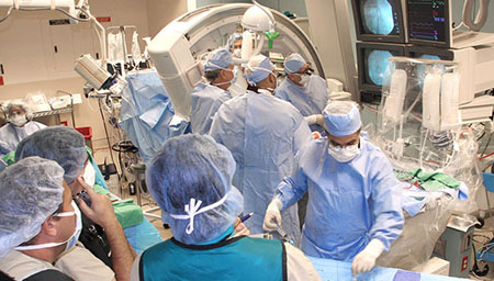
A Review of Neurosurgical Malpractice Lawsuits
We conducted a retrospective review of 287 closed neurosurgical malpractice lawsuits from February 2006 to December 2016. Data were collected on patient demographics, clinical event, timing and reason for lawsuit (misdiagnosis, delayed diagnosis/delayed treatment, unnecessary surgery, faulty surgical technique, improper postoperative care, inadequate follow-up, surgical infection, lack of informed consent), subspecialty (spinal surgery, cranial surgery, peripheral nerve surgery), operative course and legal outcome (settled or defended, out-of-court or in-court, payout where settled). There were 153 male plaintiffs and 134 female plaintiffs, and their average age was 49.6. In 79 percent of the cases patient care originated with an office visit. The vast majority of cases involved elective spine surgery for degenerative spine disease, and the majority of those spine cases involved placement of instrumentation. Among the surgical procedures, 54% were lumbosacral, 27% were cervical, 15% were cranial, and only 4% were thoracic.
The most common causes of lawsuits were faulty surgical technique (46%), misdiagnosis (10%), delayed diagnosis/delayed treatment (9%), unnecessary surgery (10%) and improper postoperative (9%) and it was lowest against surgical infection (8%), lack of informed consent (6%) and inadequate follow-up (2%). For all lawsuits, continued pain, permanent nerve damage, paralysis, infection, and need for additional surgery were the most common complaints.
The defendant was successful in 127 cases, with success defined as case dismissal, abandonment by the plaintiff, verdict for defendant, or summary judgment for the defendant. One hundred and forty-one cases were settled. A plaintiff verdict was rendered in 19 cases and a defense verdict in 27 cases.
It is not uncommon for settlements to occur in neurosurgical malpractice cases where the plaintiff has neurological deficits, despite the common belief that the deficits are unrelated to actions of the defendant. In some cases there may be an impetus to settle simply because of the severity of the plaintiff’s neurological deficits and the resulting sympathy engendered in jurists during a trial.
Neurosurgery cases pay the most to claimants of any specialty. The average payment for cases that settled was $379,860, and the average award for cases tried was $405,350. The highest median payouts were for faulty surgical technique ($430,000) and delayed diagnosis/misdiagnosis ($410,650).
Despite the advances in the field of neurosurgery over the past several decades, neurosurgery tops the list of specialties with malpractice claims made against it owing to the risk of morbidity and mortality from both operative intervention and the natural history of the clinical conditions treated. The proportion of neurosurgeons dealing with a claim each year is 19%, compared with 5% in family medicine. Because the average time to resolution of a claim is 22.3 months and the mean annual probability of a malpractice claim is 19%, it has been estimated that nearly 11 years of a surgeon’s career is spent with a malpractice claim outstanding.
Understanding the factors associated with negligence lawsuits is vital if we are to identify areas of underperformance and subsequently improve patient safety. Neurosurgeons should become familiar with the most common origins of malpractice lawsuits in the specialty. In spinal surgery, these common causes include lumbar laminectomies for discetomies, posterior cervical laminectomies, and lumbar decompression for stenosis. When performing these procedures, neurosurgeons should take extraordinary care to practice meticulous surgical technique and good patient management.
Wrong level – Minimizing medicolegal risk in spinal surgery must evidently focus on reducing faulty surgical technique, given its high incidence, high likelihood of success and potential costs. Of the successful lawsuits against faulty surgical technique in spinal surgery, thirteen were due to wrong level surgery secondary to inadequate use of intraoperative x-ray guidance to check the vertebral level. Wrong-level disc surgery is the second most common reason for reoperation for a herniated disc (recurrent disc herniation being the most common reason). Reasons for wrong-level disc surgery include:
- No localizing radiographs or fluoroscopy obtained
- Misinterpretation of localizing studies because of congenital variation (i.e. transitional anatomy), inadequate radiographic exposure or incorrect counting
- Inadequate radiographic visualization because of large body habitus or operating table limitations
- Failure to recognize that operated level does not demonstrate the expected pathology
The standard of care in spinal surgery includes operating at the correct site of the lesion using imaging guidance. Surgical technique did not meet this standard in the cases of wrong level spinal surgery and resulted directly in postoperative neurological deficits in these patients.
One successful lawsuit against faulty surgical technique due to wrong level surgery was filed two years following the procedure. In this case, the verdict highlighted the deficient recording of the patient’s preoperative neurological deficits in the medical notes, which meant that the neurosurgeon could not construct an adequate defense when explaining postoperative outcomes. Given that lawsuits may occur years after the clinical event, we emphasize the importance of thorough contemporaneous documentation in reducing the risk of future litigation.
Delayed or wrong diagnosis was the second most common lawsuit against spinal procedures. The neurosurgical facility ultimately treating the patient was not always implicated but rather the referring local hospital to which the patient first presented. In the context of clinical emergencies, delay in diagnosis or referral from the local hospital presents a high risk to patient safety and can be associated with costly litigation.
A case in our review illustrates this point. A 54-year-old patient presented to a local hospital with a 5-week history of lower back pain and bilateral leg weakness, which had worsened over the previous 48 hours to the point where she was no longer able to walk. She did not have any urinary symptoms. Although the emergency department physician considered a diagnosis of cauda equina syndrome, magnetic resonance imaging (MRI) to confirm the diagnosis was only requested on an urgent rather than emergent basis, resulting in a 24-hour delay in performing the scan. Following the imaging, there was a further 24-hour delay in radiological review and referral to the neurosurgical facility for surgery. During the combined 48 hours of delay, the patient developed established urinary retention and worsened leg weakness. Following decompression surgery for a large central L3/4 disc prolapse at the neurosurgical facility, she had a limited recovery in bladder function and has been receiving complex treatment to improve this for the last three years.
The settlement relating to this case was $1,750,000 against the local hospital. In clinical emergencies with a level of diagnostic uncertainty (e.g., cauda equina syndrome), immediate MRI to confirm the diagnosis and early consultation with a neurosurgeon are crucial to ensure timely referral for surgical intervention, and therefore also to ensure quality care and reduced risk of litigation.
Cauda equina syndrome has an incidence of 2.2 percent to 3.2 percent following lumbar disc herniation. As a postoperative complication, cauda equina syndrome is a rare problem, with an occurrence of 0.002 percent to 0.3 percent after lumbar spine surgery. In a review of 10 lawsuits with a postoperative cauda equina syndrome, six were associated with a cerebral spinal fluid leak and four were associated with an epidural hematoma. Eight of the 10 cases had a settlement or jury verdict for the plaintiff.
This syndrome is the basis for medical liability claims for several reasons. Failure to recognize the syndrome and failure to act promptly are commonly cited. A thorough history and physical exam, including documentation of rectal tone and peri-anal sensation, are crucial to making the diagnosis. Additionally, nursing staff charged with performing neurologic exams are responsible for reporting changes in motor or visceral functions and incontinence. Patients at risk of cauda equina syndrome who complain of worsening pain should be asked about their bladder function and examined if the diagnosis is suspected.
New neurologic deficit – During lumbar surgery, the incidence of motor root damage resulting in paresis occurred in 8 cases, with the L5 nerve root being the most commonly injured root. There were six lawsuits of nerve root injury resulting in foot drop and one resulting in loss of plantar flexion. Three of these cases had an associated dural tear. Six of the lawsuits were resolved in favor of the plaintiff.
With regard to the cervical spine, new neurologic deficits after cervical spine surgery are very diverse in presentation, ranging from complete quadriplegia to injury to the recurrent laryngeal nerve and to the cervical sympathetic chain. Other common reasons for suits involving the cervical spine included delay or failure to diagnose, lack of informed consent, and use of a device that had not yet received approval from the Food and Drug Administration.
Dural tear – Incidental tear of the dural sac and subsequent cerebrospinal fluid leak is possibly the most frequent intraoperative complication of lumbar spine surgery. The incidence is estimated to be 0.3 percent to 13 percent, rising to 17.6 percent with revision procedures. Although incidental durotomy is considered a benign occurrence, a subset of patients with intraoperative dural tears will develop serious problems such as pseudomeningocele, CSF fistula formation, meningitis and arachnoiditis, with new or worsened neurologic deficits or chronic pain. Intraoperative or postoperative identification of a CSF leak often results in wound healing complications, lumbar drain placement, and/or reoperation. These complications usually extend a patient’s hospital stay, can be painful, and have their own associated risks.
When a lumbar drain is placed for treatment of a spinal CSF leak, the patient should remain flat in bed. Any patient with post-dural injury headache that intensifies after an initial plateau, persists for longer than a week, or loses its orthostatic character should be evaluated for intracranial sinus or venous thrombosis.
We highlight the case of a 40-year-old woman who was diagnosed with a CSF leak after spinal surgery. Fifteen hours after placement of a lumbar drain she developed pure alexia and color agnosia caused by left lateral sinus thrombosis with hemorrhagic infarction in the posterior inferior left temporal lobe. Plaintiff’s expert was able to establish the association of intracranial venous thrombosis (IVT) with injury to the spinal dura, and the mechanism whereby the lumbar drain may facilitate its development.
Although rarely the sole basis for a lawsuit, incidental durotomies with associated sequelae appear to be a common factor prompting lawsuits. At the time of surgery, the surgeon should repair any apparent durotomy, if possible. The risk of durotomy and its potential sequelae should be discussed with the patient during the consent process. The patient should be notified of the occurrence and carefully monitored and treated for any sequelae.
Vascular and visceral injuries – Vascular and visceral injuries secondary to perforation of the anterior annulus fibrosis represent some of the most lethal complications that the neurosurgeon encounters. Vascular injuries (aorta, vena cava or iliac vessels) are estimated to occur at a rate of 1.6 to 17 per 10,000 cases, with an associated mortality of 15 percent to 62 percent. Additionally, visceral injuries are estimated to occur in 3.8 of 10,000 cases. These include injuries to the bowel, ureter, bladder and pancreas. In a review of 21 malpractice claims (18 vascular and 3 visceral injuries), the plaintiff was successful nearly half of the time.
Surgeons are often unaware that they have breached the anterior annulus or that there has been a vascular or visceral injury, because the telltale brisk bleeding associated with most large vessel injuries often does not occur. Unexplained bleeding or hypotension should raise suspicion of a vascular or visceral injury. Patients typically complain of abdominal pain and distention developing over the course of several days. If the surgeon suspects such an injury, immediate steps should be taken to obtain diagnostic studies or intra-abdominal exploration. Imaging with plain upright chest radiographs or abdominal computed tomography may demonstrate free air in the abdominal cavity.
In an example of one such case, a 45-year-old female complained of abdominal tenderness and distention immediately after microdiscectomy for L4-5 and L5-S1 disc herniation. Abdominal computed tomography revealed several foci of air overlying the anterior aspect of the vertebral body at the L5-S1 level. Segmental resection of the small bowel including small tears and primary anastomosis of the jejunum were performed.
Despite the ominous nature of these injuries, their occurrence is not considered ipso facto evidence of negligence. The standard of care with these injuries rests with the recognition and treatment of the complication rather than the occurrence itself.
Some controversy exists over informed consent with regard to these injuries. Some surgeons consider these injuries so rare that they do not warrant inclusion in the consent process other than in the general discussion of death. Other surgeons feel that it is much easier to explain an unplanned abdominal incision to the patient if the potential complication was mentioned preoperatively.
Cranial surgery There were 23 lawsuits involving cranial neurosurgical procedures (craniotomy for aneurysm clipping, craniotomy for tumor and AVM resection, brain biopsy, cranioplasty, shunt placement, craniotomy/burr holes for trauma, deep brain stimulation, and transphenoidal/transfrontal pituitary tumor resection).
The following cases illustrate two successful lawsuits against faulty surgical technique in cranial surgery: misplaced drain in brain cortex during evacuation of a subdural hematoma, resulting in a non-reversible neurological deficit, and incomplete meningioma removal. In the latter case, immediate postoperative imaging demonstrated more than 60% of residual tumor and the patient’s preoperative neurological deficit had not resolved. ‘Redo’ craniotomy and resection of the residual meningioma was undertaken, which led to a protracted hospital stay complicated by wound infection and hospital acquired pneumonia. It was conceded by the neurosurgeon that intraoperative imaging had not been utilized, and that this represented inappropriate practice, falling short of the expected standard of care, and resulted directly in a misplaced craniotomy flap that was inadequate for good tumor access and resection. The settlement for this lawsuit was $575,000.
Infection – The rate of claims against post-operative surgical infections was 5% in spinal surgery compared with 3% in cranial surgery. Craniotomy increased the risk of a shunt becoming infected. Shunt infection was more common after repeated shunt operations in quick succession. Antibiotic prophylaxis should be used not only in shunt operations but in all operations performed on patients with a shunt. If bacteria are recovered in a suspected shunt infection, immediate removal of the shunt is the best treatment. However, if the shunt’s removal or replacement is exceptionally difficult intraventricular antibiotic treatment may be tried. Clinical signs and conventional laboratory tests, apart from bacterial culture, cannot differentiate between bacterial and aseptic meningitis, but a drop in the level of consciousness suggests bacterial meningitis.
Bone flap infections accounted for more than half of all infections after supratentorial craniotomy. Bacterial meningitis accounted for more than half of all infections after suboccipital craniotomy and translabyrinthine operations. In these patients bacterial meningitis was six times more common and aseptic meningitis three times more common than in those who had had supratentorial operations.
The risk factors for infection were:
- Implantation of foreign body (associated with one-half of the infection)
- CSF leakage
- Previous neurosurgical infection
- Absence of antibiotic prophylaxis
- Duration of surgery over 4 h
- Interventions involving nasal sinuses
- Emergency surgeries
- Prior radiation therapy
Retained surgical sponges – In the medicolegal realm, a retained surgical sponge is a classic example of res ipsa loquitur (the injury speaks for itself), with the majority of cases ruled or settled in favor of the plaintiff. In our analysis, four of five cases were settled for the plaintiff.
Perioperative ischemic optic neuropathy (POION) – Despite its relative rarity, perioperative vision loss after spine surgery garners considerable interest in the medicolegal forum. Reports list an incidence of up to 0.12 percent for spine surgery; POION is more common after surgery in the prone position. Additional risk factors are intraoperative hypotension, anemia and a prolonged procedure. Recent authors have recommended that all patients undergoing spine surgery should be informed about the low but definite risk of this devastating and untreatable complication.
Peripheral nerve surgery – Peripheral nerve surgery was performed most commonly for carpal tunnel syndrome or ulnar neuropathy. There were four lawsuits against peripheral nerve surgery. The lower incidence of claims in this subspecialty compared with cranial and spinal surgery may reflect the relatively lower complexity of operative management involved and the lower attendant morbidity. As a result, patients are less likely to suffer adverse outcomes and pursue legal action.
Choosing Wisely Initiative – A continuing trend of ‘defensive medicine’ has been observed among neurosurgeons whereby clinically unnecessary diagnostic imaging and consultations may be conducted and high risk procedures may be avoided to satisfy a perceived legal risk.
As part of the Choosing Wisely initiative, the American Association of Neurological Surgeons (AANS) and Congress of Neurological Surgeons (CNS) have released a list of the 5 tests or procedures that are commonly ordered, but not always necessary, in neurosurgery.
The following 5 “evidence-based recommendations” can support physicians in working with their patients to make wise choices about their care, the organizations say:
- Don’t administer steroids after severe traumatic brain injury.
- Don’t obtain imaging (plain radiography, MRI, computed tomography [CT], or other advanced imaging) of the spine in patients with nonspecific acute low back pain and without red flags.
- Don’t routinely obtain CT scanning of children with mild head injuries.
- Don’t routinely screen for brain aneurysms in asymptomatic patients without a family or personal history of brain aneurysms, subarachnoid hemorrhage (SAH), or genetic disorders that may predispose to aneurysm formation.
- Don’t routinely use seizure prophylaxis in patients following ischemic stroke.
“Neurosurgeons are committed to identifying the right treatment, for the right patient, at the right time to help eliminate unnecessary procedures, optimize outcomes, and reduce healthcare costs,” AANS President, Robert E. Harbaugh, MD, said in a statement. “Participating in the Choosing Wisely initiative is a key step in this process.”
“There is an awful lot of waste in the medical care system,” Dr. Harbaugh said. “At least from my experience, much of that waste is treating things that don’t need to be treated, screening examinations that are unlikely to give you any meaningful information, and just unnecessary testing. Some of it is driven by the concerns for medical liability and practicing defensive medicine,” he noted.
At Top NeuroDocs, we believe that improving patient safety in neurosurgical practice requires careful attention to differential diagnosis, consideration of all relevant clinical data, thorough patient consultation, meticulous surgical technique, active pursuit of good physician-patient relationships, careful attention to the perioperative period, and adequate monitoring of patients receiving nonsurgical treatment.
The consent process should be improved to better help patients understand the possible outcomes of surgery. Whenever possible, the surgeon should obtain an appropriate and complete informed consent in the office prior to surgery, not in the hospital room or preoperative holding area, and document the consent process in the office notes. Recent studies show the importance of thorough patient consultation not only preoperatively with informed consent, to guide surgical decision making and reduce patient anxiety, but especially postoperatively, to enable patients to participate effectively in their own recovery. In all settings, thorough consultation with patients and their families is critical in neurosurgery to achieve good patient care and to reduce injury and liability.
The complications of a neurosurgery error can be devastating, including permanent brain damage, paralysis, coma, and death. Those who survive a brain surgery error may be faced with six-figure medical bills, months or years of rehabilitation, and long-term disabilities.
There’s no way to tell how often doctors lie to protect their colleagues, but ProPublica has found that patients are frequently not told the truth when they are harmed. Studies also show that many physicians do not have a favorable view of informing patients about mistakes and health care workers are afraid to speak up if things don’t seem right. Many doctors and nurses have told ProPublica that they fear retaliation if they speak out about patient safety problems.
Effective communication is vital; physicians should develop a clear, straightforward style of communicating with patients, colleagues and other members of the treatment team. If a mistake does occur, tell the patient directly and express sincere empathy and concern. Patients appreciate this type of candor, and expressing sympathy is never an admission of malpractice.
Thorough documentation is also important. It is not a good idea to rely on memory rather than documentation. Neurosurgeons should document every case completely, including their thought processes and all relevant details. Records should be organized and legible. Good documentation can make all the difference if a lawsuit is filed.
Leave a Reply
Want to join the discussion?Feel free to contribute!